Contents
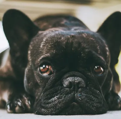
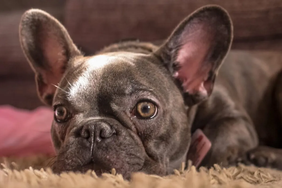
French Bulldogs are predisposed to a range of eye conditions that most other breeds encounter far less frequently. The anatomy that makes them appealing, those large, round, expressive eyes, also makes those same eyes structurally vulnerable. Understanding what to watch for and when to act can prevent conditions that start as minor irritations from progressing into serious problems.
This guide covers the most significant eye conditions seen in the breed. For cherry eye specifically, there is a dedicated cherry eye article and a surgery cost guide.
Why Frenchie eyes need extra attention
The brachycephalic skull compresses the facial anatomy. In French Bulldogs, this means the eye sockets are relatively shallow, causing the eyes to protrude beyond the normal level of protection offered by the orbital rim. The exposed eyeball is more susceptible to:
- Direct trauma (scratches, foreign bodies, contact with vegetation)
- Corneal dryness due to incomplete blinking
- Friction from facial skin folds around the eye socket
- Hair from nasal folds rubbing against the corneal surface
This is not speculation: VetCompass data from the Royal Veterinary College consistently shows French Bulldogs to have significantly higher rates of corneal ulcers, conjunctivitis and lid abnormalities than the general dog population.
Routine eye checks at home, part of a regular grooming and handling routine, catch early problems before they become serious.
Cherry eye (prolapsed nictitans gland)
Cherry eye is one of the most recognisable eye conditions in the breed. It presents as a round, red or pink mass in the inner corner of one or both eyes, produced by the gland of the third eyelid prolapsing outward from its normal position.
The third eyelid (nictitating membrane) sits in the inner corner of the eye and provides supplementary protection and a significant proportion of the eye’s tear production. When the connective tissue anchoring the gland weakens, the gland flips outward and becomes visible.
Cherry eye requires treatment. If left uncorrected, the exposed gland becomes inflamed, prone to infection and gradually loses its ability to produce tears, increasing the risk of dry eye (KCS) later in life.
The preferred surgical treatment is the pocket technique, in which the gland is tucked back into place rather than removed. Preserving the gland maintains its contribution to the tear film. Removal of the gland was previously more common but is now considered suboptimal unless pocket surgery has failed.
Detailed surgical guidance and costs are in the dedicated cherry eye surgery guide.
Corneal ulcers
A corneal ulcer is a break in the surface layer (epithelium) of the cornea. In Frenchies, the most common causes are:
- Trauma from nasal fold hair or skin rubbing the eye surface
- Foreign bodies: a grass seed, piece of grit or fragment of something the dog has been digging in
- Inadequate blink coverage due to the prominent eye shape
- Secondary to dry eye, where the dried surface breaks down
Signs of a corneal ulcer: the dog holds the eye partially closed, there is redness of the eye and surrounding tissue, increased discharge, tearing, and the dog may paw at the face. In fluorescein staining at the vet, the dye lights up the defect.
Superficial ulcers typically respond well to antibiotic and lubricating eye drops over five to seven days. Deeper ulcers require more intensive treatment and monitoring. Deep stromal ulcers, where the defect has penetrated a significant depth into the cornea, can progress to perforation and are surgical emergencies.
A Frenchie with any signs of eye pain or discomfort should be seen by a vet the same day, not the following morning.
Dry eye (keratoconjunctivitis sicca)
Dry eye occurs when the tear glands produce insufficient tears to maintain a healthy corneal surface. Without adequate lubrication, the cornea dries, becomes irritated, and eventually suffers damage including pigmentation, clouding and ulceration.
Signs include:
- Red, inflamed-looking eyes
- A thick, greenish or yellow-grey discharge (rather than the watery discharge of a normal eye)
- The dog blinking frequently or squinting
- Dull, cloudy appearance to the corneal surface
Dry eye is diagnosed by a Schirmer Tear Test (a small paper strip placed in the eye for a minute to measure tear production). A reading below 15mm per minute is considered abnormal; below 10mm is diagnostic for KCS.
Management is usually lifelong: cyclosporine or tacrolimus eye drops stimulate the tear glands to produce more tears in many cases, and lubricating eye drops maintain the surface between doses. The condition is controllable rather than curable in most dogs.
The connection to cherry eye is important: if the nictitans gland was removed (rather than repositioned) during cherry eye surgery, the reduced tear production can trigger or worsen dry eye. This is one of the main reasons the pocket technique is now preferred.
Entropion
Entropion is the inward rolling of one or both eyelids, causing the eyelashes and skin to continuously rub against the corneal surface. It is painful, produces chronic discharge and squinting, and causes progressive corneal damage including pigmentation and scarring if untreated.
In French Bulldogs, entropion most commonly affects the lower lid at the outer corner of the eye, though it can occur at the inner corner or the upper lid. It may be present from a young age or develop as the dog matures and facial skin laxity changes.
Mild entropion in puppies may be managed with lubrication and monitoring while the face is still growing. Established entropion in adult dogs requires surgical correction: a small amount of skin is removed from the eyelid to pull it back to its correct position.
Distichiasis
Distichiasis is the presence of extra eyelashes (distichiae) growing from abnormal positions on the eyelid margin. These extra lashes rub against the corneal surface with every blink.
Many dogs with distichiasis are not significantly affected. When the extra lashes cause corneal irritation, discharge or ulceration, treatment is needed. Options include manual removal (temporary, as the lashes regrow), electrolysis (permanent destruction of the follicle) or cryotherapy.
French Bulldogs are over-represented in distichiasis diagnoses, likely related to the same anatomical predispositions that affect other parts of their ocular anatomy.
Hereditary cataracts
The Kennel Club recommends DNA testing of breeding French Bulldogs for hereditary cataracts (HC-HSF4). The DNA test identifies whether a dog carries zero, one or two copies of the mutation associated with early-onset hereditary cataracts.
Dogs that are DNA Clear (no copies of the mutation) will not develop the hereditary form and cannot pass it to offspring. Carriers (one copy) will not develop the condition themselves but can pass the mutation to offspring. Affected dogs (two copies) have a high likelihood of developing cataracts.
This is one of the reasons responsible breeders DNA test their breeding dogs and one of the health test results you should ask to see when buying a puppy.
Pigmentary keratitis
Pigmentary keratitis (PK) is the deposition of dark pigment on the corneal surface, often starting at the nasal side of the eye. It typically develops secondary to chronic irritation: from entropion, nasal fold hair rubbing, inadequate blink coverage or dry eye.
As the pigment spreads across the cornea, it reduces vision. Severe, central pigmentary keratitis can cause significant visual impairment.
Treatment addresses the underlying cause where possible (surgical correction of entropion, nasal fold reduction, management of dry eye) and may include cyclosporine drops, which have modest evidence for slowing or reducing pigment deposition. This is an area where early intervention in underlying conditions pays dividends long-term.
Nasal fold skin and the eye surface
The nasal fold that runs from the nose up toward the inner corner of the eye is present to varying degrees in French Bulldogs. In some dogs, the fold is tight enough that the hair growing on it makes direct contact with the corneal surface of the medial (inner) eye.
Checking regularly whether the nasal fold hair is touching the eye is part of routine home care. If it is, a vet or groomer can trim the hair. In severe cases, surgical reduction of the nasal fold is considered. The grooming guide covers routine home checks.
When to call the vet
Contact your vet the same day if your Frenchie shows:
- Holding an eye partially or fully closed
- Sudden or significant increase in discharge
- Visible redness of the white of the eye or the tissue around the eye
- Pawing or rubbing at the eye or face
- A visible mass in the inner corner of the eye (cherry eye)
- Apparent cloudiness or change in the corneal surface appearance
- Squinting
Overnight or weekend: if you cannot reach your vet and the dog is showing signs of acute eye pain, an emergency veterinary service should be consulted. Corneal injuries that penetrate deeply deteriorate quickly.
The earlier eye problems are identified and treated, the better the outcome. Most eye conditions in French Bulldogs respond well when caught early and deteriorate significantly when left. For the cosmetic but common issue of reddish-brown staining below the eyes, its causes, the safe removal methods and when it requires investigation, the French Bulldog tear stains guide covers the full picture.
Frequently asked questions
-
The French Bulldog's flat face means the eye sockets are shallower and the eyes protrude more than in dogs with normal skull anatomy. This exposure makes the cornea (the clear surface of the eye) more vulnerable to injury, dryness and infection. The same anatomy reduces the blink reflex's effectiveness and leaves the tear film less evenly distributed. Additionally, the excess facial skin folds around the eye can rub the surface, and the prominent eyes are more likely to trap hair and debris.
-
Cherry eye is the prolapse of the third eyelid gland (nictitans gland). French Bulldogs have a third eyelid in the inner corner of each eye that contains a tear-producing gland. When the connective tissue holding the gland in place weakens, the gland prolapses outward and appears as a round, pink or red mass in the inner corner of the eye. It typically requires surgical correction, either a pocket technique (preferred) or, in some cases, removal of the gland.
-
Keratoconjunctivitis sicca (KCS), known as dry eye, is a condition in which insufficient tear production leads to chronic dryness of the corneal surface. The eyes appear red and uncomfortable, with a thick, sticky discharge. Left untreated, it causes progressive corneal damage, clouding and ulceration. Dry eye in Frenchies can be a long-term consequence of cherry eye treatment (particularly if the third eyelid gland was removed) and is managed with daily eye drops.
-
Corneal ulcers range from superficial and straightforward to treat, to deep and potentially sight-threatening. Because Frenchie eyes protrude and are exposed to friction from skin folds, they are at higher risk of corneal damage. Any eye that is held partially closed, is red, is producing discharge, or where the dog is pawing at their face should be seen by a vet the same day. Untreated corneal ulcers deepen and can lead to perforation in severe cases.
-
Entropion is a condition in which the eyelid rolls inward, causing the eyelashes and skin to rub against the corneal surface. It produces chronic irritation, squinting, discharge and corneal damage over time. Entropion can be congenital (present from birth) or develop with age. Mild entropion may be managed medically in puppies to allow growth before surgical correction is considered. Established entropion typically requires surgical correction.
-
Eye conditions are generally covered by good pet insurance policies provided they are not pre-existing conditions at the time the policy is taken out. Lifetime policies are essential for conditions like dry eye or hereditary cataracts that require ongoing treatment. Cherry eye, entropion and corneal ulcer treatment are typically covered as acute or surgically correctable conditions. Always read the exclusions carefully; some policies exclude conditions linked to the breed's anatomy.