Contents
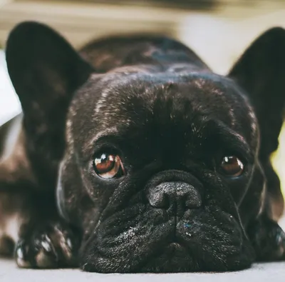
Cherry eye is one of the most recognisable eye conditions in French Bulldogs. The sudden appearance of a red or pink mass in the inner corner of the eye is alarming the first time you see it, but it is a well-understood condition with effective treatment when addressed promptly. This guide explains what cherry eye is, why French Bulldogs are prone to it, what treatment looks like and what to expect after surgery.
If you are looking specifically for UK cost information, see the cherry eye surgery cost guide. For the broader picture of eye conditions in the breed, the eye problems guide covers dry eye, corneal ulcers, entropion and other conditions common in brachycephalic dogs.
What cherry eye is
Dogs, unlike humans, have a third eyelid: the nictitating membrane, which sits in the inner corner of each eye and can sweep across the eye surface for additional protection and lubrication. Within this third eyelid sits a gland, the nictitans gland (or gland of the third eyelid), that produces a significant portion of the eye’s total tear film, estimates suggest around 30 to 40 per cent in dogs.
This gland is anchored to the orbital tissue by a ligament-like structure. In dogs with the connective tissue laxity associated with brachycephalic anatomy, this attachment can be weak. When the attachment fails, the gland prolapes out from behind the third eyelid and becomes visible as the characteristic red or pink swelling in the inner corner of the eye.
The condition is not painful in itself when it first occurs, though repeated friction and drying of the exposed gland causes progressive damage that becomes uncomfortable over time.
Why French Bulldogs are particularly prone to cherry eye
The flat face of a brachycephalic breed like the French Bulldog is associated with structural changes throughout the skull, including the orbit (eye socket). The shallower orbital depth and altered orbital anatomy in flat-faced dogs affects how the eye sits and how the associated structures are anchored.
French Bulldogs also tend to have prominent, less recessed eyes compared to longer-nosed breeds. This combination of orbital anatomy and the connective tissue characteristics associated with the breed increases the incidence of cherry eye significantly compared to non-brachycephalic breeds.
Cherry eye can occur at any age but is most common in dogs under two years old. It is not unusual for one eye to be affected first and the other to follow months or years later.
Recognising cherry eye
Cherry eye is usually unmistakable: a rounded, pink or red mass appearing at the inner corner of one or both eyes. It may appear suddenly and seem to come and go at first, particularly early in the condition. Over time, without treatment, it tends to remain prolapsed more consistently.
In the early stages, the affected eye may not appear uncomfortable. As the condition progresses and the gland is increasingly exposed to air and friction:
- The gland becomes swollen and inflamed
- The eye may develop discharge
- The dog may paw at the eye or show signs of discomfort
- Reduced tear production may begin as gland function is impaired
If you see the characteristic pink swelling in the corner of your Frenchie’s eye, book a vet appointment within a day or two. Early treatment gives the best long-term outcome.
What happens if cherry eye is left untreated
The gland of the third eyelid is responsible for a substantial portion of tear production. When it is prolapsed and exposed, the tissue is progressively damaged by dehydration and friction. This reduces the gland’s ability to produce tears even if it is later surgically repositioned.
The long-term risk of untreated or repeatedly untreated cherry eye is keratoconjunctivitis sicca, commonly called dry eye. Dry eye occurs when tear production falls below the level needed to keep the eye surface properly lubricated. It is a chronic, progressive condition requiring lifelong management with lubricating eye drops, and in severe cases can lead to corneal damage and vision impairment.
Treating cherry eye early, before gland damage is significant, gives the best chance of preserving the gland’s function and avoiding dry eye later in life.
Treatment options
Surgical replacement (pocket or tacking technique)
The current standard of care for cherry eye is surgical repositioning of the prolapsed gland. The two main techniques are:
The pocket technique: The surgeon creates a small pocket in the tissue of the third eyelid and tucks the gland back into its normal position within this pocket, securing it with sutures. The gland is preserved entirely and its normal position restored.
The tacking technique: The gland is sutured to the orbital tissue to anchor it back into its correct position. The approach is similar in intent to the pocket technique; the specific method varies by surgeon preference and the anatomy of the individual dog.
Both approaches aim to restore the gland to its normal position while preserving its function. Most veterinary surgeons and all specialist ophthalmologists now favour gland-preserving techniques over removal.
Gland removal
Historically, cherry eye was sometimes treated by simply removing the prolapsed gland. This approach is now strongly discouraged by veterinary ophthalmology bodies because the permanent reduction in tear production it causes significantly increases the lifetime risk of dry eye.
Gland removal should only be considered in cases where the gland is too severely damaged to function and where multiple repositioning attempts have failed. It is not an appropriate first-line treatment.
Temporary management
Some owners, pending surgery or seeking a second opinion, use lubricating eye drops to keep the exposed gland moist and reduce friction damage. Manual replacement (gently pushing the gland back into position) has been described anecdotally but is not a sustainable treatment and can cause additional trauma. These approaches are at best a short-term measure while arranging surgical treatment.
UK surgery costs
Cherry eye surgery at a general practice using the pocket or tacking technique typically costs £300 to £700 per eye. If both eyes require treatment in one procedure, costs may be reduced. Referral to a veterinary ophthalmologist costs more: typically £800 to £1,500 per eye.
Recurrent cases are often referred to an ophthalmologist for a specialist assessment and a higher-success repair. A detailed breakdown of costs, insurance considerations and what to expect at consultation is in the cherry eye surgery cost guide.
Most comprehensive pet insurance policies cover cherry eye surgery as a non-pre-existing condition. Starting insurance before any eye problems present is the best way to ensure this is covered.
After surgery
Recovery from pocket or tacking technique surgery is generally straightforward. Your vet will typically prescribe:
- A course of antibiotic eye drops or ointment
- Anti-inflammatory medication
- An Elizabethan collar (cone) for one to two weeks to prevent the dog pawing at the eye
Most dogs are comfortable within 24 to 48 hours of surgery. The cone is the main source of frustration during recovery. Keeping the eye clean with prescribed drops and preventing self-trauma are the most important aftercare tasks.
A follow-up check at two to four weeks confirms the repair is holding. The recurrence rate for gland repositioning varies from around 5 to 20 per cent. If the gland prolapses again after surgery, a second repositioning attempt is preferable to removal.
Preventing cherry eye
There is no proven preventive measure. Cherry eye is a conformation-related condition associated with the brachycephalic anatomy that French Bulldogs have structurally. Health testing of breeding dogs reduces the frequency of the condition in offspring: responsible breeders will know the eye health history of their lines and will avoid pairing dogs with strong eye problem histories.
For routine eye care, including daily cleaning of the eye area and what to look for during regular health checks, see the grooming guide.
Frequently asked questions
-
Cherry eye is the common name for prolapse of the gland of the third eyelid (nictitans gland). Dogs have a third eyelid in each eye (the nictitating membrane) that contains a gland responsible for producing a significant portion of the eye's tear film. In some dogs, particularly brachycephalic breeds, the connective tissue that anchors this gland is weak, and the gland can prolapse out from behind the third eyelid, appearing as a red or pink swelling in the inner corner of the eye. The appearance gives the condition its common name.
-
Cherry eye is one of the more common ocular conditions in the breed. Brachycephalic dogs are at elevated risk because the shallow orbital socket and changes in orbital anatomy associated with the flat face affect how the gland sits and is anchored. Not every Frenchie will develop cherry eye, but it is common enough that every owner should know how to recognise it. Some dogs develop it in one eye; some develop it in both, either simultaneously or sequentially.
-
Yes. Cherry eye that is left untreated exposes the gland to repeated friction, drying and trauma, which damages the gland tissue over time and reduces its tear-producing function. Untreated or repeatedly prolapsing cherry eye increases the long-term risk of keratoconjunctivitis sicca (dry eye), a chronic condition that requires lifelong management. Early surgical treatment gives the best outcome for long-term eye health.
-
The replacement approach (also called the pocket or tacking technique) aims to reposition the prolapsed gland back into its normal position and secure it there. Because the gland is preserved, its tear-producing function is maintained. This is now the strongly preferred approach in veterinary ophthalmology. Gland removal was more commonly performed historically but is now discouraged because removing the gland permanently reduces tear production and increases the lifetime risk of dry eye. Most veterinary ophthalmologists and many general practices now offer gland replacement as standard.
-
Cherry eye surgery costs in the UK typically range from £300 to £700 per eye for the pocket or tacking technique at a general practice. Referral to a veterinary ophthalmologist (recommended for recurrent cases or complex presentations) costs more, often £800 to £1,500 per eye. If both eyes require treatment, costs can be substantially higher. Most pet insurance policies with a decent per-condition limit cover cherry eye surgery.
-
Yes. The recurrence rate varies depending on the surgical technique and the surgeon's experience. The pocket technique has a recurrence rate of roughly 5 to 20 per cent in the literature, though this varies considerably. If cherry eye recurs after the first surgery, a second attempt at repositioning is generally preferred to removal. In cases of repeated recurrence, referral to a veterinary ophthalmologist for a specialist assessment gives the best chance of a successful repair.